
Question: Vertebrate Histology Please Answer Each Activity And Question In Great Detail For Understanding. Vertebrate Histology Please Answer Each Activity And Question In Great Detail.
Vertebrate Histology
Please answer each activity and question in great detail forunderstanding.
Vertebrate Histology
please answer each activity and question in greatdetail.
Transcribed Image Text from this Question
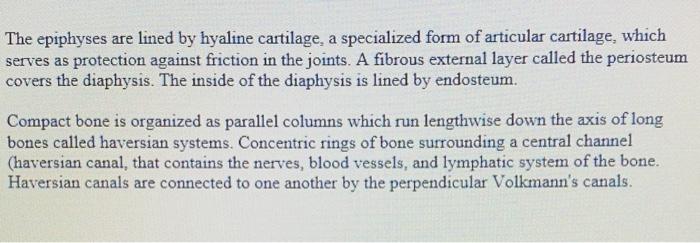
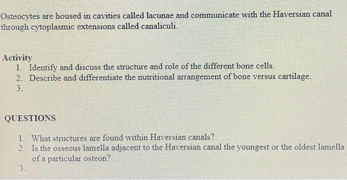
Learning Objectives 1. Describe the processes of bone remodeling and bone growth. 2. Name the different histological regions of bone. 3. Describe the calcification processes of cartilage and bone. Introduction The skeletal system is composed of bone and cartilage and functions in the protection of vital organs, mechanical support for sites of muscle attachment, storage of calcium and phosphate ions. Like all connective tissue, the components of bone are cells and matrix. Three cell types (osteoblasts, osteocytes, and osteoclasts) function in the formation and maintenance of bone. Osteoblasts synthesize the bone matrix and are responsible for its mineralization. Osteocytes are inactive osteoblasts that have become trapped within the bone they have formed. Osteoclasts, which are derived from monocytes, break down bone matrix through phagocytosis. The area between osteoclast and bone is known as Howship’s lacuna. Bone matrix is composed of osteoid which is the unmineralized matrix composed of type I collagen and glycosaminoglycans (GAGs). Calcium hydroxyapatite is a calcium salt crystal that gives bone its strength and rigidity Bone is grouped into two types based on structural and functional differences. Compact bone is thick and dense has a mechanical function. It is the area of bone to which ligaments and tendons attach Trabecular bone (spongy) bone is located in between layers of compact bone and is thin and porous. It houses the bone marrow Long bones, composed of both compact and spongy bone, contain the epiphyses at their articular surface (joint). The diaphysis is the shaft of the bone and has walls of compact bone and a network of trabecular bone. The epiphyseal growth plate lies at the region between the shaft and the epiphysis. The metaphysis is the region where the shaft of the bone joins the epiphyseal growth plate The epiphyses are lined by hyaline cartilage, a specialized form of articular cartilage, which serves as protection against friction in the joints. A fibrous external layer called the periosteum covers the diaphysis. The inside of the diaphysis is lined by endosteum. Compact bone is organized as parallel columns which run lengthwise down the axis of long bones called haversian systems. Concentric rings of bone surrounding a central channel (haversian canal, that contains the nerves, blood vessels, and lymphatic system of the bone. Haversian canals are connected to one another by the perpendicular Volkmann’s canals. Osteocytes are housed in cavities called lacunae and communicate with the Haversian canal through cytoplasmic extensions called canaliculi. Activity 1. Identify and discuss the structure and role of the different bone cells. 2. Describe and differentiate the nutritional arrangement of bone versus cartilage. 3. | QUESTIONS 1. What structures are found within Haversian canals? 2. Is the osseous lamella adjacent to the Haversian canal the youngest or the oldest lamella of a particular osteon? 3. Introduction The skeletal system is composed of bone and cartilage and functions in the protection of vital organs, mechanical support for sites of muscle attachment, storage of calcium and phosphate ions. Like all connective tissue, the components of bone are cells and matrix. Three cell types (osteoblasts, osteocytes, and osteoclasts) function in the formation and maintenance of bone. Osteoblasts synthesize the bone matrix and are responsible for its mineralization. Osteocytes are inactive osteoblasts that have become trapped within the bone they have formed. Osteoclasts, which are derived from monocytes, break down bone matrix through phagocytosis. The area between osteoclast and bone is known as Howship’s lacuna. Bone matrix is composed of osteoid which is the unmineralized matrix composed of type I collagen and glycosaminoglycans (GAGs). Calcium hydroxyapatite is a calcium salt crystal that gives bone its strength and rigidity. Bone is grouped into two types based on structural and functional differences. Compact bone is thick and dense has a mechanical function. It is the area of bone to which ligaments and tendons attach Trabecular bone (spongy) bone is located in between layers of compact bone and is thin and porous. It houses the bone marrow. Long bones, composed of both compact and spongy bone, contain the epiphyses at their articular surface (joint). The diaphysis is the shaft of the bone and has walls of compact bone and a network of trabecular bone. The epiphyseal growth plate lies at the region between the shaft and the epiphysis. The metaphysis is the region where the shaft of the bone joins the epiphyseal growth plate. The epiphyses are lined by hyaline cartilage, a specialized form of articular cartilage, which serves as protection against friction in the joints. A fibrous external layer called the periosteum covers the diaphysis. The inside of the diaphysis is lined by endosteum. Compact bone is organized as parallel columns which run lengthwise down the axis of long bones called haversian systems. Concentric rings of bone surrounding a central channel (haversian canal, that contains the nerves, blood vessels, and lymphatic system of the bone. Haversian canals are connected to one another by the perpendicular Volkmann’s canals. Osteocytes are housed in cavities called lacunae and communicate with the Haversian canal through cytoplasmic extensions called canaliculi. Activity 1. Identify and discuss the structure and role of the different bone cells. 2. Describe and differentiate the nutritional arrangement of bone versus cartilage. 3. QUESTIONS 1. What structures are found within Haversian canals? 2. Is the osseous lamella adjacent to the Haversian canal the youngest or the oldest lamella of a particular osteon? 3.
